参考文献
[1] Zhang X, Guo Z, Zhu L, et al. Challenges and chances coexist: A visualized analysis and bibliometric study of research on bioresorbable vascular scaffolds from 2000 to 2022[J]. Medicine (Baltimore), 2023, 102(21):e33885. DOI: 10.1097/MD.0000000000033885.
[2] Sotomi Y, Onuma Y, Miyazaki Y, et al. Is quantitative coronary angiography reliable in assessing the late lumen loss of the everolimus‑eluting bioresorbable polylactide scaffold in comparison with the cobalt‑chromium metallic stent?[J]. EuroIntervention, 2017, 13(5):e585‑e594. DOI: 10.4244/EIJ‑D‑17‑00070.
[3] Sotomi Y, Onuma Y, Suwannasom P, et al. Is quantitative coronary angiography reliable in assessing the lumen gain after treatment with the everolimus‑eluting bioresorbable polylactide scaffold?[J]. EuroIntervention, 2016, 12(8):e998‑e1008. DOI: 10.4244/EIJV12I8A163.
[4] Gomez‑Lara J, Brugaletta S, Farooq V, et al. Angiographic geometric changes of the lumen arterial wall after bioresorbable vascular scaffolds and metallic platform stents at 1‑year follow‑up[J]. JACC Cardiovasc Interv, 2011, 4(7):789‑799. DOI: 10.1016/j.jcin.2011.04.009.
[5] Nallamothu BK, Spertus JA, Lansky AJ, et al. Comparison of clinical interpretation with visual assessment and quantitative coronary angiography in patients undergoing percutaneous coronary intervention in contemporary practice: the Assessing Angiography (A2) project[J]. Circulation, 2013, 127(17):1793‑1800. DOI: 10.1161/CIRCULATIONAHA.113.001952.
[6] Gutiérrez‑Chico JL, Serruys PW, Girasis C, et al. Quantitative multi‑modality imaging analysis of a fully bioresorbable stent: a head‑to‑head comparison between QCA, IVUS and OCT[J]. Int J Cardiovasc Imaging, 2012, 28(3):467‑478. DOI: 10.1007/s10554‑011‑9829‑y.
[7] Witberg G, Lavi I, Vaknin Assa H, et al. Insights from 2D and 3D quantitative angiographic assessment of bioresorbable everolimus‑eluting vascular scaffolds[J]. Isr Med Assoc J, 2016, 18(7):318‑385.
[8] 中华医学会心血管病学分会.冠状动脉生物可吸收支架临床应用中国专家共识[J].中华心血管病杂志,2020,48(5):350‑358. DOI:10.3760/cma.j.cn112148‑20200317‑00224.
[9] Onuma Y, Honda Y, Asano T, et al. Randomized comparison between everolimus‑eluting bioresorbable scaffold and metallic stent: multimodality imaging through 3 years[J]. JACC Cardiovasc Interv, 2020, 13(1):116‑127. DOI: 10.1016/j.jcin.2019.09.047.
[10] Han Y, Xu B, Fu G, et al. A randomized trial comparing the neovas sirolimus‑eluting bioresorbable scaffold and metallic everolimus‑eluting stents[J]. JACC Cardiovasc Interv, 2018, 11(3):260‑272. DOI: 10.1016/j.jcin.2017.09.037.
[11] Gao R, Yang Y, Han Y, et al. Bioresorbable vascular scaffolds versus metallic stents in patients with coronary artery disease: ABSORB China Trial[J]. J Am Coll Cardiol, 2015, 66(21):2298‑2309. DOI: 10.1016/j.jacc.2015.09.054.
[12] 武玙璠, 汤超杰, 王武.生物可吸收血管内支架的现状[J].介入放射学杂志,2023, 32(10): 1040‑1049. DOI: 10.3969/j.issn.1008‑794X.2023.10.20.
[13] Sotomi Y, Suwannasom P, Tenekecioglu E, et al. Differential aspects between cobalt‑chromium everolimus drug‑eluting stent and Absorb everolimus bioresorbable vascular scaffold: from bench to clinical use[J]. Expert Rev Cardiovasc Ther, 2015, 13(10):1127‑1145. DOI: 10.1586/14779072.2015.1089172.
[14] Campos CM, Ishibashi Y, Eggermont J, et al. Echogenicity as a surrogate for bioresorbable everolimus‑eluting scaffold degradation: analysis at 1‑, 3‑, 6‑, 12‑18, 24‑, 30‑, 36‑ and 42‑month follow‑up in a porcine model[J]. Int J Cardiovasc Imaging, 2015, 31(3):471‑482. DOI: 10.1007/s10554‑015‑0591‑4.
[15] Ferdous MM, Jie Z, Gao L, et al. A first‑in‑human study of the bioheart sirolimus‑eluting bioresorbable vascular scaffold in patients with coronary artery disease: Two‑year clinical and imaging outcomes[J]. Adv Ther, 2022, 39(8):3749‑3765. DOI: 10.1007/s12325‑022‑02154‑w.
[16] Zhang YJ, Wang XZ, Fu G, et al. Clinical and multimodality imaging results at 6 months of a bioresorbable sirolimus‑eluting scaffold for patients with single de novo coronary artery lesions: the NeoVas first‑in‑man trial[J]. EuroIntervention, 2016, 12(10):1279‑1287. DOI: 10.4244/EIJV12I10A209.
[17] Serruys PW, Onuma Y, Ormiston JA, et al. Evaluation of the second generation of a bioresorbable everolimus drug‑eluting vascular scaffold for treatment of de novo coronary artery stenosis: six‑month clinical and imaging outcomes[J]. Circulation, 2010, 122(22):2301‑2312. DOI: 10.1161/CIRCULATIONAHA.110.970772.
[18] Serruys PW, Onuma Y, Garcia‑Garcia HM, et al. Dynamics of vessel wall changes following the implantation of the absorb everolimus‑eluting bioresorbable vascular scaffold: a multi‑imaging modality study at 6, 12, 24 and 36 months[J]. EuroIntervention, 2014, 9(11):1271‑1284. DOI: 10.4244/EIJV9I11A217.
[19] van Zandvoort L, Dudek D, Weber‑Albers J, et al. Intravascular ultrasound findings of the Fantom sirolimus‑eluting bioresorbable scaffold at six‑and nine‑month follow‑up: the FANTOM II study[J]. EuroIntervention, 2018, 14(11):e1215‑e1223. DOI: 10.4244/EIJ‑D‑18‑00491.
[20] Mintz GS, Nissen SE, Anderson WD, et al. American College of Cardiology clinical expert consensus document on standards for acquisition, Measurement and reporting of intravascular ultrasound studies (IVUS). A report of the American College of Cardiology Task Force on Clinical Expert Consensus Documents[J]. J Am Coll Cardiol, 2001, 37(5):1478‑1492. DOI: 10.1016/s0735‑1097(01)01175‑5.
[21] Okada K, Honda Y, Kitahara H, et al. Bioresorbable scaffold for treatment of coronary artery lesions: intravascular ultrasound results from the ABSORB Japan trial[J]. JACC Cardiovasc Interv, 2018, 11(7):648‑661. DOI: 10.1016/j.jcin.2017.11.034.
[22] Nishi T, Okada K, Kitahara H, et al. Intravascular ultrasound predictors of long‑term outcomes following ABSORB bioresorbable scaffold implantation: A pooled analysis of the ABSORB III and ABSORB Japan trials[J]. J Cardiol, 2021, 78(3):224‑229. DOI: 10.1016/j.jjcc.2021.03.005.
[23] Okada K, Honda Y, Kitahara H, et al. Scaffold underexpansion and late lumen loss after bioresorbable scaffold implantation: Insights from ABSORB JAPAN trial[J]. Int J Cardiol Heart Vasc, 2020,31:100623. DOI: 10.1016/j.ijcha.2020.100623.
[24] Kawamoto H, Ruparelia N, Latib A, et al. Expansion in calcific lesions and overall clinical outcomes following bioresorbable scaffold implantation optimized with intravascular ultrasound[J]. Catheter Cardiovasc Interv, 2017, 89(5):789‑797. DOI: 10.1002/ccd.26725.
[25] Shan P, Motivala A, Moses J, et al. Imaging comparison of a bioresorbable vascular scaffold by high‑frequency intravascular ultrasound and optical coherence tomography[J]. JACC Cardiovasc Interv, 2015, 8(11):e187‑e188. DOI: 10.1016/j.jcin.2015.05.023.
[26] Okada K, Kitahara H, Mitsutake Y, et al. Assessment of bioresorbable scaffold with a novel high‑definition 60 MHz IVUS imaging system: Comparison with 40MHz IVUS referenced to optical coherence tomography[J]. Catheter Cardiovasc Interv, 2018, 91(5):874‑883. DOI: 10.1002/ccd.27197.
[27] Munding CE, Chérin E, Alves N, et al. 30/80 MHz Bidirectional dual‑frequency IVUS feasibility evaluated in vivo and for stent imaging[J]. Ultrasound Med Biol, 2020, 46(8):2104‑2112. DOI: 10.1016/j.ultrasmedbio.2020.03.032.
[28] Wu Y, Shen L, Yin J, et al. 5 years of serial intravascular imaging outcomes of XINSORB sirolimus‑eluting bioresorbable vascular scaffold[J]. JACC Cardiovasc Interv, 2019, 12(6):602‑603. DOI: 10.1016/j.jcin.2018.11.029.
[29] Ono M, Kawashima H, Hara H, et al. Advances in IVUS/OCT and future clinical perspective of novel hybrid catheter system in coronary imaging[J]. Front Cardiovasc Med, 2020,7:119. DOI: 10.3389/fcvm.2020.00119.
[30] Brugaletta S, Garcia‑Garcia HM, Diletti R, et al. Comparison between the first and second generation bioresorbable vascular scaffolds: a six month virtual histology study[J]. EuroIntervention, 2011, 6(9):1110‑1116. DOI: 10.4244/EIJV6I9A193.
[31] Zeng Y, Tateishi H, Cavalcante R, et al. Serial assessment of tissue precursors and progression of coronary calcification analyzed by fusion of IVUS and OCT: 5‑year follow‑up of scaffolded and nonscaffolded arteries[J]. JACC Cardiovasc Imaging, 2017, 10(10 Pt A):1151‑1161. DOI: 10.1016/j.jcmg.2016.11.016.
[32] Brugaletta S, Garcia‑Garcia HM, Serruys PW, et al. NIRS and IVUS for characterization of atherosclerosis in patients undergoing coronary angiography[J]. JACC Cardiovasc Imaging, 2011, 4(6):647‑655. DOI: 10.1016/j.jcmg.2011.03.013.
[33] Terada K, Kubo T, Kameyama T, et al. NIRS‑IVUS for differentiating coronary plaque rupture, erosion, and calcified nodule in acute myocardial infarction[J]. JACC Cardiovasc Imaging, 2021, 14(7):1440‑1450. DOI: 10.1016/j.jcmg.2020.08.030.
[34] Noori M, Thayssen P, Veien KT, et al. Lipid‑core burden response to stent implantation assessed with near‑infrared spectroscopy and intravascular ultrasound evaluation in patients with myocardial infarction[J]. Cardiovasc Revasc Med, 2017, 18(3):182‑189. DOI: 10.1016/j.carrev.2016.12.018.
[35] Roleder T, Dobrolinska M, Pociask E, et al. Coronary plaque redistribution after stent implantation is determined by lipid composition: A NIRS‑IVUS analysis[J]. Cardiol J, 2020, 27(3):238‑245. DOI: 10.5603/CJ.a2018.0111.
[36] Stone GW, Maehara A, Ali ZA, et al. Percutaneous coronary intervention for vulnerable coronary atherosclerotic plaque[J]. J Am Coll Cardiol, 2020, 76(20):2289‑2301. DOI: 10.1016/j.jacc.2020.09.547.
[37] de Graaf FR, Schuijf JD, van Velzen JE, et al. Diagnostic accuracy of 320‑row multidetector computed tomography coronary angiography to noninvasively assess in‑stent restenosis[J]. Invest Radiol, 2010, 45(6):331‑340. DOI: 10.1097/RLI.0b013e3181dfa312.
[38] Nagayama Y, Emoto T, Hayashi H, et al. Coronary stent evaluation by CTA: Image quality comparison between super‑resolution deep learning reconstruction and other reconstruction algorithms[J]. AJR Am J Roentgenol, 2023, 221(5):599‑610. DOI: 10.2214/AJR.23.29506.
[39] Onuma Y, Dudek D, Thuesen L, et al. Five‑year clinical and functional multislice computed tomography angiographic results after coronary implantation of the fully resorbable polymeric everolimus‑eluting scaffold in patients with de novo coronary artery disease: the ABSORB cohort A trial[J]. JACC Cardiovasc Interv, 2013, 6(10):999‑1009. DOI: 10.1016/j.jcin.2013.05.017.
[40] Collet C, Chevalier B, Cequier A, et al. Diagnostic accuracy of coronary CT angiography for the evaluation of bioresorbable vascular scaffolds[J]. JACC Cardiovasc Imaging, 2018, 11(5):722‑732. DOI: 10.1016/j.jcmg.2017.04.013.
[41] Collet C, Sotomi Y, Cavalcante R, et al. Accuracy of coronary computed tomography angiography for bioresorbable scaffold luminal investigation: a comparison with optical coherence tomography[J]. Int J Cardiovasc Imaging, 2017, 33(3):431‑439. DOI: 10.1007/s10554‑016‑1018‑6.
[42] Ghafari C, Brassart N, Delmotte P, et al. Bioresorbable magnesium‑based stent: real‑world clinical experience and feasibility of follow‑up by coronary computed tomography: a new window to look at new scaffolds[J]. Biomedicines, 2023, 11(4): 1150. DOI: 10.3390/biomedicines11041150.
[43] Reiss S, Krafft AJ, Zehender M, et al. Magnetic resonance imaging of bioresorbable vascular scaffolds: potential approach for noninvasive evaluation of coronary patency[J]. Circ Cardiovasc Interv, 2015, 8(4):e002388.DOI: 10.1161/CIRCINTERVENTIONS.115.002388.
[44] Aoki J, Kishi S, Saito S, et al. Coronary magnetic resonance angiographic images of a lesion treated with a metal stent and fully bioresorbable polymeric everolimus‑eluting scaffold[J]. EuroIntervention, 2016, 12(12):1519. DOI: 10.4244/EIJ‑D‑16‑00263.
[45] Barone‑Rochette G, Vautrin E, Rodière M, et al. First magnetic resonance coronary artery imaging of bioresorbable vascular scaffold in‑patient[J]. Eur Heart J Cardiovasc Imaging, 2015, 16(2):229. DOI: 10.1093/ehjci/jeu203.
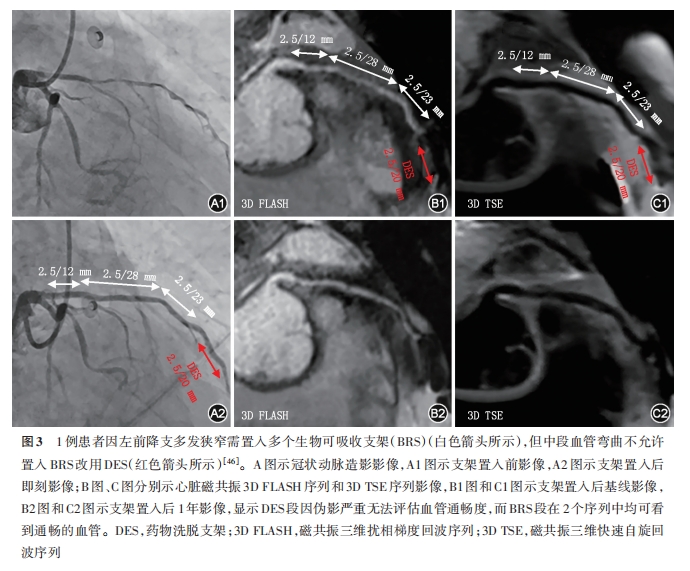
[46] von Zur Mühlen C, Reiss S, Krafft AJ, et al. Coronary magnetic resonance imaging after routine implantation of bioresorbable vascular scaffolds allows non‑invasive evaluation of vascular patency[J]. PLoS One, 2018, 13(1):e0191413. DOI: 10.1371/journal.pone.0191413.
[47] Bian D, Qin L, Lin W, et al. Magnetic resonance (MR) safety and compatibility of a novel iron bioresorbable scaffold[J]. Bioact Mater, 2020, 5(2):260‑274. DOI: 10.1016/j.bioactmat.2020.02.011.
[48] Lawton JS, Tamis‑Holland JE, Bangalore S, et al. 2021 ACC/AHA/SCAI guideline for coronary artery revascularization: Executive summary: A report of the American College of Cardiology/American Heart Association joint committee on clinical practice guidelines[J]. Circulation, 2022, 145(3):e4‑e17. DOI: 10.1161/CIR.0000000000001039.
[49] Sedlis SP, Lorin JD. Should fractional flow reserve be measured after stent deployment? Routinely? Ever?[J]. JACC Cardiovasc Interv, 2017, 10(10):996‑998. DOI: 10.1016/j.jcin.2017.03.020.
[50] van de Hoef TP, Siebes M, Spaan JA, et al. Fundamentals in clinical coronary physiology: why coronary flow is more important than coronary pressure[J]. Eur Heart J, 2015, 36(47):3312‑3319a. DOI: 10.1093/eurheartj/ehv235.
[51] Tonino PA, De Bruyne B, Pijls NH, et al. Fractional flow reserve versus angiography for guiding percutaneous coronary intervention[J]. N Engl J Med, 2009, 360(3):213‑224. DOI: 10.1056/NEJMoa0807611.
[52] Tu S, Barbato E, Köszegi Z, et al. Fractional flow reserve calculation from 3‑dimensional quantitative coronary angiography and TIMI frame count: a fast computer model to quantify the functional significance of moderately obstructed coronary arteries[J]. JACC Cardiovasc Interv, 2014, 7(7):768‑777. DOI: 10.1016/j.jcin.2014.03.004.
[53] Tu S, Westra J, Yang J, et al. Diagnostic accuracy of fast computational approaches to derive fractional flow reserve from diagnostic coronary angiography: The international multicenter FAVOR pilot study[J]. JACC Cardiovasc Interv, 2016, 9(19):2024‑2035. DOI: 10.1016/j.jcin.2016.07.013.
[54] Li Z, Zhan J, Han J, et al. Comparison of diagnostic accuracy of immediate angiography derived residual quantitative flow ratio after bioresorbable scaffold and drug eluting stent implantation[J]. Rev Cardiovasc Med, 2022, 23(2):59. DOI: 10.31083/j.rcm2302059.
[55] Saito Y, Cristea E, Bouras G, et al. Long‑term serial functional evaluation after implantation of the Fantom sirolimus‑eluting bioresorbable coronary scaffold[J]. Catheter Cardiovasc Interv, 2021, 97(3):431‑436. DOI: 10.1002/ccd.28804.
[56] Asano T, Katagiri Y, Collet C, et al. Functional comparison between the BuMA Supreme biodegradable polymer sirolimus‑eluting stent and a durable polymer zotarolimus‑eluting coronary stent using quantitative flow ratio: PIONEER QFR substudy[J]. EuroIntervention, 2018, 14(5):e570‑e579. DOI: 10.4244/EIJ‑D‑17‑00461.
[57] Chen J, Wetzel LH, Pope KL, et al. FFRCT: Current status[J]. AJR Am J Roentgenol, 2021, 216(3):640‑648. DOI: 10.2214/AJR.20.23332.
[58] Zhuang B, Wang S, Zhao S, et al. Computed tomography angiography‑derived fractional flow reserve (CT‑FFR) for the detection of myocardial ischemia with invasive fractional flow reserve as reference: systematic review and meta‑analysis[J]. Eur Radiol, 2020, 30(2):712‑725. DOI: 10.1007/s00330‑019‑06470‑8.
[59] Tang CX, Guo BJ, Schoepf JU, et al. Feasibility and prognostic role of machine learning‑based FFRCT in patients with stent implantation[J]. Eur Radiol, 2021, 31(9):6592‑6604. DOI: 10.1007/s00330‑021‑07922‑w.
[60] Pontone G, Andreini D, Guaricci AI, et al. Incremental diagnostic value of stress computed tomography myocardial perfusion with whole‑heart coverage CT scanner in intermediate‑to high‑risk symptomatic patients suspected of coronary artery disease[J]. JACC Cardiovasc Imaging, 2019, 12(2):338‑349. DOI: 10.1016/j.jcmg.2017.10.025.
[61] Andreini D, Mushtaq S, Pontone G, et al. Rationale and design of advantage (additional diagnostic value of CT perfusion over coronary CT angiography in stented patients with suspected in‑stent restenosis or coronary artery disease progression) prospective study[J]. J Cardiovasc Comput Tomogr, 2018, 12(5):411‑417. DOI: 10.1016/j.jcct.2018.06.003.
[62] Andreini D, Mushtaq S, Pontone G, et al. CT perfusion versus coronary CT angiography in patients with suspected in‑stent restenosis or CAD progression[J]. JACC Cardiovasc Imaging, 2020, 13(3):732‑742. DOI: 10.1016/j.jcmg.2019.05.031.
[63] Stuijfzand WJ, Schumacher SP, Driessen RS, et al. Myocardial blood flow and coronary flow reserve during 3 years following bioresorbable vascular scaffold versus metallic drug‑eluting stent implantation: the VANISH trial[J]. JACC Cardiovasc Interv, 2019, 12(10):967‑979. DOI: 10.1016/j.jcin.2019.03.004.
[64] Gutiérrez‑Chico JL, Chen Y, Yu W, et al. Diagnostic accuracy and reproducibility of optical flow ratio for functional evaluation of coronary stenosis in a prospective series[J]. Cardiol J, 2020, 27(4):350‑361. DOI: 10.5603/CJ.a2020.0071.
[65] Huang J, Emori H, Ding D, et al. Diagnostic performance of intracoronary optical coherence tomography‑based versus angiography‑based fractional flow reserve for the evaluation of coronary lesions[J]. EuroIntervention, 2020, 16(7):568‑576. DOI: 10.4244/EIJ‑D‑19‑01034.
[66] Emori H, Kubo T, Shiono Y, et al. Comparison of optical flow ratio and fractional flow ratio in stent‑treated arteries immediately after percutaneous coronary intervention[J]. Circ J, 2020, 84(12):2253‑2258. DOI: 10.1253/circj.CJ‑20‑0661.
[67] Ding D, Yu W, Tauzin H, et al. Optical flow ratio for assessing stenting result and physiological significance of residual disease[J]. EuroIntervention, 2021, 17(12):e989‑e998. DOI: 10.4244/EIJ‑D‑21‑00185.
[68] Yu W, Tanigaki T, Ding D, et al. Accuracy of intravascular ultrasound‑based fractional flow reserve in identifying hemodynamic significance of coronary stenosis[J]. Circ Cardiovasc Interv, 2021, 14(2):e009840. DOI: 10.1161/CIRCINTERVENTIONS.120.009840.
[69] Yong D, Minjie C, Yujie Z, et al. Diagnostic performance of IVUS‑FFR analysis based on generative adversarial network and bifurcation fractal law for assessing myocardial ischemia[J]. Front Cardiovasc Med, 2023,10:1155969. DOI: 10.3389/fcvm.2023.1155969.
[70] Bezerra CG, Hideo‑Kajita A, Bulant CA, et al. Coronary fractional flow reserve derived from intravascular ultrasound imaging: Validation of a new computational method of fusion between anatomy and physiology[J]. Catheter Cardiovasc Interv, 2019, 93(2):266‑274. DOI: 10.1002/ccd.27822.
[71] Sui Y, Yang M, Xu Y, et al. Diagnostic performance of intravascular ultrasound‑based fractional flow reserve versus angiography‑based quantitative flow ratio measurements for evaluating left main coronary artery stenosis[J]. Catheter Cardiovasc Interv, 2022, 99 Suppl 1∶1403‑1409. DOI: 10.1002/ccd.30078.
[72] Lin W, Zhang H, Zhang W, et al. In vivo degradation and endothelialization of an iron bioresorbable scaffold[J]. Bioact Mater, 2021, 6(4):1028‑1039. DOI: 10.1016/j.bioactmat.2020.09.020.
[73] Wang L, Zhao Y, Zheng B, et al. Ultrawide‑bandwidth high‑resolution all‑optical intravascular ultrasound using miniaturized photoacoustic transducer[J]. Sci Adv, 2023, 9(23):eadg8600. DOI: 10.1126/sciadv.adg8600.
[74] Lau YS, Tan LK, Chan CK, et al. Automated segmentation of metal stent and bioresorbable vascular scaffold in intravascular optical coherence tomography images using deep learning architectures[J]. Phys Med Biol, 2021, 66(24):5026. DOI: 10.1088/1361‑6560/ac4348.
[75] Tu S, Holm NR, Koning G, et al. Fusion of 3D QCA and IVUS/OCT[J]. Int J Cardiovasc Imaging, 2011, 27(2):197‑207. DOI: 10.1007/s10554‑011‑9809‑2.
[76] Poon E, Wu X, Dijkstra J, et al. Angiography and optical coherence tomography derived shear stress: are they equivalent in my opinion?[J]. Int J Cardiovasc Imaging, 2023, 39(10):1953‑1961. DOI: 10.1007/s10554‑023‑02949‑0.
[77] Wu W, Khan B, Sharzehee M, et al. Three dimensional reconstruction of coronary artery stents from optical coherence tomography: experimental validation and clinical feasibility[J]. Sci Rep, 2021, 11(1):12252. DOI: 10.1038/s41598‑021‑91458‑y.