参考文献
[1] Qaseem A, Wilt TJ, Rich R, et al. Pharmacologic treatment of hypertension in adults aged 60 years or older to higher versus lower blood pressure targets: a clinical practice guideline from the American College of Physicians and the American Academy of Family Physicians[J]. Ann Intern Med, 2017,166(6):430‑437. DOI: 10.7326/M16‑1785.
[2] Williams B, Mancia G, Spiering W, et al. 2018 ESC/ESH Guidelines for the management of arterial hypertension. The Task Force for the management of arterial hypertension of the European Society of Cardiology (ESC) and the European Society of Hypertension (ESH)[J]. G Ital Cardiol (Rome), 2018,19(11 Suppl 1):3S‑73S. DOI: 10.1714/3026.30245.
[3] Whelton PK, Carey RM, Aronow WS, et al. 2017 ACC/AHA/AAPA/ABC/ACPM/AGS/APhA/ASH/ASPC/NMA/PCNA guideline for the prevention, detection, evaluation, and management of high blood pressure in adults: a report of the American College of Cardiology/American Heart Association Task Force on clinical practice guidelines[J]. J Am Coll Cardiol, 2018,71(19):e127‑e248. DOI: 10.1016/j.jacc.2017.11.006.
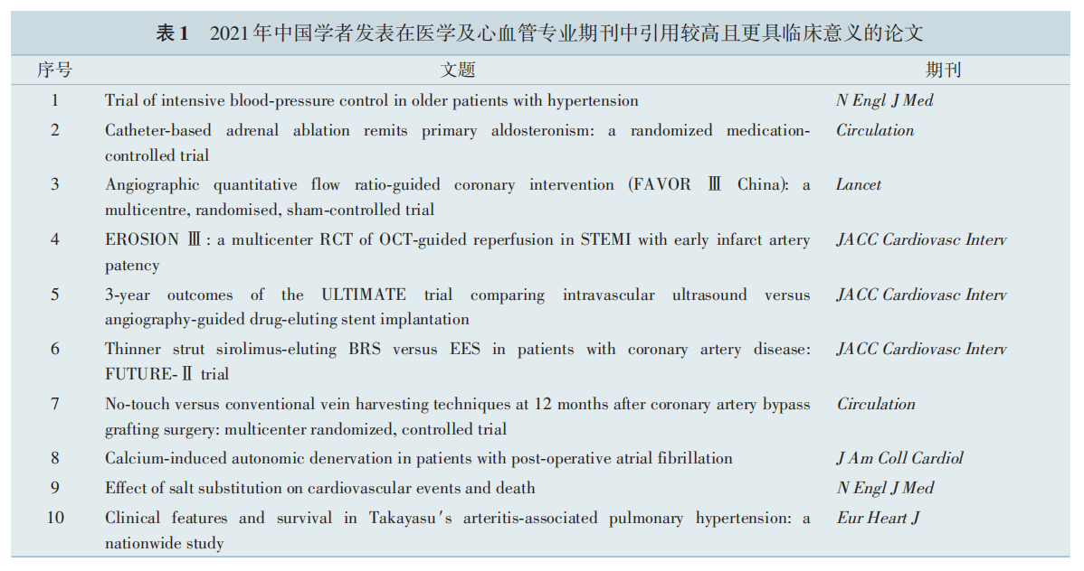
[4] Zhang W, Zhang S, Deng Y, et al. Trial of intensive blood‑pressure control in older patients with hypertension[J]. N Engl J Med,2021,385(14):1268‑1279. DOI: 10.1056/NEJMoa2111437.
[5] SPRINT Research Group, Lewis CE, Fine LJ, et al. Final Report of a Trial of Intensive versus Standard Blood‑Pressure Control[J]. N Engl J Med, 2021,384(20):1921‑1930. DOI: 10.1056/NEJMoa1901281.
[6] Wang A, Tian X, Zuo Y, et al. Control of blood pressure and risk of cardiovascular disease and mortality in elderly chinese: a real‑world prospective cohort study[J]. Hypertension,2022,79(8):1866‑1875. DOI: 10.1161/HYPERTENSIONAHA.122.19587.
[7] Sacks BA, Sacks AC, Faintuch S. Radiofrequency ablation treatment for aldosterone‑producing adenomas[J]. Curr Opin Endocrinol Diabetes Obes, 2017,24(3):169‑173. DOI: 10.1097/MED.0000000000000329.
[8] Fowler AM, Burda JF, Kim SK. Adrenal artery embolization: anatomy, indications, and technical considerations[J]. AJR Am J Roentgenol, 2013,201(1):190‑201. DOI: 10.2214/AJR.12.9507.
[9] Zhao Z, Liu X, Zhang H, et al. Catheter‑based adrenal ablation remits primary aldosteronism: a randomized medication‑controlled trial[J]. Circulation, 2021,144(7):580‑582. DOI: 10.1161/CIRCULATIONAHA.121.054318.
[10] Zhang H, Li Q, Liu X, et al. Adrenal artery ablation for primary aldosteronism without apparent aldosteronoma: An efficacy and safety, proof‑of‑principle trial[J]. J Clin Hypertens (Greenwich), 2020,22(9):1618‑1626. DOI: 10.1111/jch.13960.
[11] Park SJ, Kang SJ, Ahn JM, et al. Visual‑functional mismatch between coronary angiography and fractional flow reserve[J]. JACC Cardiovasc Interv, 2012,5(10):1029‑1036. DOI: 10.1016/j.jcin.2012.07.007.
[12] Toth G, Hamilos M, Pyxaras S, et al. Evolving concepts of angiogram: fractional flow reserve discordances in 4000 coronary stenoses[J]. Eur Heart J, 2014,35(40):2831‑2838. DOI: 10.1093/eurheartj/ehu094.
[13] van Nunen LX, Zimmermann FM, Tonino PA, et al. Fractional flow reserve versus angiography for guidance of PCI in patients with multivessel coronary artery disease (FAME): 5‑year follow‑up of a randomised controlled trial[J]. Lancet, 2015,386(10006):1853‑1860. DOI: 10.1016/S0140‑6736(15)00057‑4.
[14] Tonino PA, De Bruyne B, Pijls NH, et al. Fractional flow reserve versus angiography for guiding percutaneous coronary intervention[J]. N Engl J Med, 2009,360(3):213‑224. DOI: 10.1056/NEJMoa0807611.
[15] Neumann FJ, Sousa‑Uva M, Ahlsson A, et al. 2018 ESC/EACTS Guidelines on myocardial revascularization. The Task Force on myocardial revascularization of the European Society of Cardiology (ESC) and European Association for Cardio‑Thoracic Surgery (EACTS)[J]. G Ital Cardiol (Rome), 2019,20(7‑8 Suppl 1):1S‑61S. DOI: 10.1714/3203.31801.
[16] Li J, Gong Y, Wang W, et al. Accuracy of computational pressure‑fluid dynamics applied to coronary angiography to derive fractional flow reserve: FLASH FFR[J]. Cardiovasc Res, 2020,116(7):1349‑1356. DOI: 10.1093/cvr/cvz289.
[17] Tang CX, Liu CY, Lu MJ, et al. CT FFR for ischemia‑specific CAD with a new computational fluid dynamics algorithm: a Chinese multicenter study[J]. JACC Cardiovasc Imaging, 2020,13(4):980‑990. DOI: 10.1016/j.jcmg.2019.06.018.
[18] Götberg M, Berntorp K, Rylance R, et al. 5-year outcomes of PCI guided by measurement of instantaneous wave-free ratio versus fractional flow reserve[J]. J Am Coll Cardiol,2022,79(10):965-974. DOI: 10.1016/j.jacc.2021.12.030.
[19] Tu S, Westra J, Yang J, et al. Diagnostic accuracy of fast computational approaches to derive fractional flow reserve from diagnostic coronary angiography: the international multicenter FAVOR pilot study[J]. JACC Cardiovasc Interv, 2016,9(19):2024‑2035. DOI: 10.1016/j.jcin.2016.07.013.
[20] Xu B, Tu S, Song L, et al. Angiographic quantitative flow ratio‑guided coronary intervention (FAVOR III China): a multicentre, randomised, sham‑controlled trial[J]. Lancet, 2021,398(10317):2149‑2159. DOI: 10.1016/S0140‑6736(21)02248‑0.
[21] Jia H, Dai J, Hou J, et al. Effective anti‑thrombotic therapy without stenting: intravascular optical coherence tomography‑based management in plaque erosion (the EROSION study)[J]. Eur Heart J,2017,38(11):792‑800. DOI: 10.1093/eurheartj/ehw381.
[22] Jia H, Dai J, He L, et al. EROSION III: a multicenter RCT of OCT‑guided reperfusion in stemi with early infarct artery patency[J]. JACC Cardiovasc Interv, 2022,15(8):846‑856. DOI: 10.1016/j.jcin.2022.01.298.
[23] Wijns W, Shite J, Jones MR, et al. Optical coherence tomography imaging during percutaneous coronary intervention impacts physician decision‑making: ILUMIEN I study[J]. Eur Heart J, 2015,36(47):3346‑3355. DOI: 10.1093/eurheartj/ehv367.
[24] Khalifa A, Kubo T, Shimamura K, et al. Impact of optical coherence tomography imaging on decision‑making during percutaneous coronary intervention in patients presented with acute coronary syndromes[J]. Circ J, 2021,85(10):1781‑1788. DOI: 10.1253/circj.CJ‑20‑0942.
[25] Kinnaird T, Johnson T, Anderson R, et al. Intravascular imaging and 12‑month mortality after unprotected left main stem PCI: an analysis from the british cardiovascular intervention society database[J]. JACC Cardiovasc Interv, 2020,13(3):346‑357. DOI: 10.1016/j.jcin.2019.10.007.
[26] Andell P, Karlsson S, Mohammad MA, et al. Intravascular ultrasound guidance is associated with better outcome in patients undergoing unprotected left main coronary artery stenting compared with angiography guidance alone[J]. Circ Cardiovasc Interv, 2017,10(5): e004813. DOI: 10.1161/CIRCINTERVENTIONS.116.004813.
[27] Kim JS, Kang TS, Mintz GS, et al. Randomized comparison of clinical outcomes between intravascular ultrasound and angiography‑guided drug‑eluting stent implantation for long coronary artery stenoses[J]. JACC Cardiovasc Interv, 2013,6(4):369‑376. DOI: 10.1016/j.jcin.2012.11.009.
[28] Tian NL, Gami SK, Ye F, et al. Angiographic and clinical comparisons of intravascular ultrasound‑versus angiography‑guided drug‑eluting stent implantation for patients with chronic total occlusion lesions: two‑year results from a randomised AIR‑CTO study[J]. EuroIntervention, 2015,10(12):1409‑1417. DOI: 10.4244/EIJV10I12A245.
[29] Chen L, Xu T, Xue XJ, et al. Intravascular ultrasound‑guided drug‑eluting stent implantation is associated with improved clinical outcomes in patients with unstable angina and complex coronary artery true bifurcation lesions[J]. Int J Cardiovasc Imaging, 2018,34(11):1685‑1696. DOI: 10.1007/s10554‑018‑1393‑2.
[30] Zhang J, Gao X, Kan J, et al. Intravascular ultrasound versus angiography‑guided drug‑eluting stent implantation: the ULTIMATE trial[J]. J Am Coll Cardiol, 2018,72(24):3126‑3137. DOI: 10.1016/j.jacc.2018.09.013.
[31] Gao XF, Ge Z, Kong XQ, et al. 3‑Year Outcomes of the ULTIMATE trial comparing intravascular ultrasound versus angiography‑guided drug‑eluting stent implantation[J]. JACC Cardiovasc Interv, 2021,14(3):247‑257. DOI: 10.1016/j.jcin.2020.10.001.
[32] Mentias A, Sarrazin MV, Saad M, et al. Long‑term outcomes of coronary stenting with and without use of intravascular ultrasound[J]. JACC Cardiovasc Interv, 2020,13(16):1880‑1890. DOI: 10.1016/j.jcin.2020.04.052.
[33] Serruys PW, Ormiston JA, Onuma Y, et al. A bioabsorbable everolimus‑eluting coronary stent system (ABSORB): 2‑year outcomes and results from multiple imaging methods[J]. Lancet, 2009,373(9667):897‑910. DOI: 10.1016/S0140‑6736(09)60325‑1.
[34] Ellis SG, Kereiakes DJ, Metzger DC, et al. Everolimus‑eluting bioresorbable scaffolds for coronary artery disease[J]. N Engl J Med, 2015,373(20):1905‑1915. DOI: 10.1056/NEJMoa1509038.
[35] Gao R, Yang Y, Han Y, et al. Bioresorbable vascular scaffolds versus metallic stents in patients with coronary artery disease: ABSORB China trial[J]. J Am Coll Cardiol, 2015,66(21):2298‑2309. DOI: 10.1016/j.jacc.2015.09.054.
[36] Kimura T, Kozuma K, Tanabe K, et al. A randomized trial evaluating everolimus‑eluting Absorb bioresorbable scaffolds vs. everolimus‑eluting metallic stents in patients with coronary artery disease: ABSORB Japan[J]. Eur Heart J, 2015,36(47):3332‑3342. DOI: 10.1093/eurheartj/ehv435.
[37] Waksman R. Bioresorbable scaffolds versus metallic stents in routine PCI[J]. N Engl J Med, 2017,377(18):1790‑1791. DOI: 10.1056/NEJMc1711903.
[38] Kereiakes DJ, Ellis SG, Metzger C, et al. 3‑year clinical outcomes with everolimus‑eluting bioresorbable coronary scaffolds: the ABSORB III trial[J]. J Am Coll Cardiol, 2017,70(23):2852‑2862. DOI: 10.1016/j.jacc.2017.10.010.
[39] Yamaji K, Ueki Y, Souteyrand G, et al. Mechanisms of very late bioresorbable scaffold thrombosis: the invest registry[J]. J Am Coll Cardiol, 2017,70(19):2330‑2344. DOI: 10.1016/j.jacc.2017.09.014.
[40] Tenekecioglu E, Torii R, Katagiri Y, et al. Early strut protrusion and late neointima thickness in the Absorb bioresorbable scaffold: a serial wall shear stress analysis up to five years[J]. EuroIntervention, 2019,15(4):e370‑e379. DOI: 10.4244/EIJ‑D‑18‑00381.
[41] Song L, Xu B, Chen Y, et al. Thinner strut sirolimus‑eluting brs versus ees in patients with coronary artery disease: FUTURE‑II Trial[J]. JACC Cardiovasc Interv, 2021,14(13):1450‑1462. DOI: 10.1016/j.jcin.2021.04.048.
[42] Han Y, Xu B, Fu G, et al. A randomized trial comparing the neovas sirolimus‑eluting bioresorbable scaffold and metallic everolimus‑eluting stents[J]. JACC Cardiovasc Interv, 2018,11(3):260‑272. DOI: 10.1016/j.jcin.2017.09.037.
[43] Harskamp RE, Lopes RD, Baisden CE, et al. Saphenous vein graft failure after coronary artery bypass surgery: pathophysiology, management, and future directions[J]. Ann Surg, 2013,257(5):824‑833. DOI: 10.1097/SLA.0b013e318288c38d.
[44] Souza DS, Dashwood MR, Tsui JC, et al. Improved patency in vein grafts harvested with surrounding tissue: results of a randomized study using three harvesting techniques[J]. Ann Thorac Surg, 2002,73(4):1189‑1195. DOI: 10.1016/s0003‑4975(02)03425‑2.
[45] Souza DS, Johansson B, Bojö L, et al. Harvesting the saphenous vein with surrounding tissue for CABG provides long‑term graft patency comparable to the left internal thoracic artery: results of a randomized longitudinal trial[J]. J Thorac Cardiovasc Surg, 2006,132(2):373‑378. DOI: 10.1016/j.jtcvs.2006.04.002.
[46] Neumann FJ, Sousa‑Uva M, Ahlsson A, et al. 2018 ESC/EACTS Guidelines on myocardial revascularization[J]. Eur Heart J, 2019,40(2):87‑165. DOI: 10.1093/eurheartj/ehy394.
[47] Tian M, Wang X, Sun H, et al. No‑touch versus conventional vein harvesting techniques at 12 months after coronary artery bypass grafting surgery: multicenter randomized, controlled trial[J]. Circulation, 2021,144(14):1120‑1129. DOI: 10.1161/CIRCULATIONAHA.121.055525.
[48] Deb S, Singh SK, de Souza D, et al. SUPERIOR SVG: no touch saphenous harvesting to improve patency following coronary bypass grafting (a multi‑Centre randomized control trial, NCT01047449)[J]. J Cardiothorac Surg, 2019,14(1):85. DOI: 10.1186/s13019‑019‑0887‑x.
[49] Filardo G, Damiano RJ Jr, Ailawadi G, et al. Epidemiology of new‑onset atrial fibrillation following coronary artery bypass graft surgery[J]. Heart, 2018,104(12):985‑992. DOI: 10.1136/heartjnl‑2017‑312150.
[50] Maesen B, Nijs J, Maessen J, et al. Post‑operative atrial fibrillation: a maze of mechanisms[J]. Europace, 2012,14(2):159‑174. DOI: 10.1093/europace/eur208.
[51] Amar D, Zhang H, Miodownik S, et al. Competing autonomic mechanisms precede the onset of postoperative atrial fibrillation[J]. J Am Coll Cardiol, 2003,42(7):1262‑1268. DOI: 10.1016/s0735‑1097(03)00955‑0.
[52] Shen MJ, Choi EK, Tan AY, et al. Neural mechanisms of atrial arrhythmias[J]. Nat Rev Cardiol, 2011,9(1):30‑39. DOI: 10.1038/nrcardio.2011.139.
[53] Patterson E, Po SS, Scherlag BJ, et al. Triggered firing in pulmonary veins initiated by in vitro autonomic nerve stimulation[J]. Heart Rhythm, 2005,2(6):624‑631. DOI: 10.1016/j.hrthm.2005.02.012.
[54] Wang H, Zhang Y, Xin F, et al. Calcium‑induced autonomic denervation in patients with post‑operative atrial fibrillation[J]. J Am Coll Cardiol, 2021,77(1):57‑67. DOI: 10.1016/j.jacc.2020.10.049.
[55] Pokushalov E, Kozlov B, Romanov A, et al. Botulinum toxin injection in epicardial fat pads can prevent recurrences of atrial fibrillation after cardiac surgery: results of a randomized pilot study[J]. J Am Coll Cardiol, 2014,64(6):628‑629. DOI: 10.1016/j.jacc.2014.04.062.
[56] Waldron NH, Cooter M, Haney JC, et al. Temporary autonomic modulation with botulinum toxin type A to reduce atrial fibrillation after cardiac surgery[J]. Heart Rhythm, 2019,16(2):178‑184. DOI: 10.1016/j.hrthm.2018.08.021.
[57] Aburto NJ, Hanson S, Gutierrez H, et al. Effect of increased potassium intake on cardiovascular risk factors and disease: systematic review and meta‑analyses[J]. BMJ, 2013,346:f1378. DOI: 10.1136/bmj.f1378.
[58] Aburto NJ, Ziolkovska A, Hooper L, et al. Effect of lower sodium intake on health: systematic review and meta‑analyses[J]. BMJ, 2013,346:f1326. DOI: 10.1136/bmj.f1326.
[59] Huang L, Trieu K, Yoshimura S, et al. Effect of dose and duration of reduction in dietary sodium on blood pressure levels: systematic review and meta‑analysis of randomised trials[J]. BMJ, 2020,368:m315. DOI: 10.1136/bmj.m315.
[60] Filippini T, Naska A, Kasdagli MI, et al. Potassium intake and blood pressure: a dose‑response meta‑analysis of randomized controlled trials[J]. J Am Heart Assoc, 2020,9(12):e015719. DOI: 10.1161/JAHA.119.015719.
[61] Greer RC, Marklund M, Anderson C, et al. Potassium‑enriched salt substitutes as a means to lower blood pressure: benefits and risks[J]. Hypertension, 2020,75(2):266‑274. DOI: 10.1161/HYPERTENSIONAHA.119.13241.
[62] Neal B, Wu Y, Feng X, et al. Effect of salt substitution on cardiovascular events and death[J]. N Engl J Med, 2021,385(12):1067‑1077. DOI: 10.1056/NEJMoa2105675.
[63] Lupi E, Sánchez G, Horwitz S, et al. Pulmonary artery involvement in Takayasu′s arteritis[J]. Chest, 1975,67(1):69‑74. DOI: 10.1378/chest.67.1.69.
[64] He Y, Lv N, Dang A, et al. Pulmonary artery involvement in patients with Takayasu arteritis[J]. J Rheumatol, 2020,47(2):264‑272. DOI: 10.3899/jrheum.190045.
[65] Kong X, Ma L, Lv P, et al. Involvement of the pulmonary arteries in patients with Takayasu arteritis: a prospective study from a single centre in China[J]. Arthritis Res Ther, 2020,22(1):131. DOI: 10.1186/s13075‑020‑02203‑1.
[66] Jiang X, Zhu YJ, Zhou YP, et al. Clinical features and survival in Takayasu′s arteritis‑associated pulmonary hypertension: a nationwide study[J]. Eur Heart J, 2021,42(42):4298‑4305. DOI: 10.1093/eurheartj/ehab599.
[67] Kerr GS, Hallahan CW, Giordano J, et al. Takayasu arteritis[J]. Ann Intern Med, 1994,120(11):919‑929. DOI: 10.7326/0003‑4819‑120‑11‑199406010‑00004.
[68] Yajima M, Numano F, Park YB, et al. Comparative studies of patients with Takayasu arteritis in Japan, Korea and India‑‑comparison of clinical manifestations, angiography and HLA‑B antigen[J]. Jpn Circ J, 1994,58(1):9‑14. DOI: 10.1253/jcj.58.9.