参考文献
[1] 马丽媛 , 吴亚哲 , 王文 , 等 . 《中国心血管病报告 2017》要点解读 [J]. 中国心血管杂志 ,2018,23(1):3-6. DOI: 10.3969/j.issn.1007-5410.2018.01.002.
[2] Kung G, Konstantinidis K, Kitsis RN. Programmed necrosis, not apoptosis, in the heart[J]. Circ Res, 2011,108(8):1017-1036. DOI: 10.1161/CIRCRESAHA.110.225730.
[3] Whelan RS, Kaplinskiy V, Kitsis RN. Cell death in the pathogenesis of heart disease: mechanisms and significance[J]. Annu Rev Physiol, 2010,72:19-44. DOI: 10.1146/annurev.physiol.010908.163111.
[4] Hausenloy DJ, Botker HE, Engstrom T, et al. Targeting reperfusion injury in patients with ST-segment elevation myocardial infarction: trials and tribulations[J]. Eur Heart J, 2017,38(13):935-941. DOI: 10.1093/eurheartj/ehw145.
[5] Heusch G, Gersh BJ. The pathophysiology of acute myocardial infarction and strategies of protection beyond reperfusion: a continual challenge[J]. Eur Heart J, 2017,38(11):774-784. DOI: 10.1093/eurheartj/ehw224.
[6] Marks AR. Calcium cycling proteins and heart failure: mechanisms and therapeutics[J]. J Clin Invest, 2013,123(1):46-52. DOI: 10.1172/JCI62834.
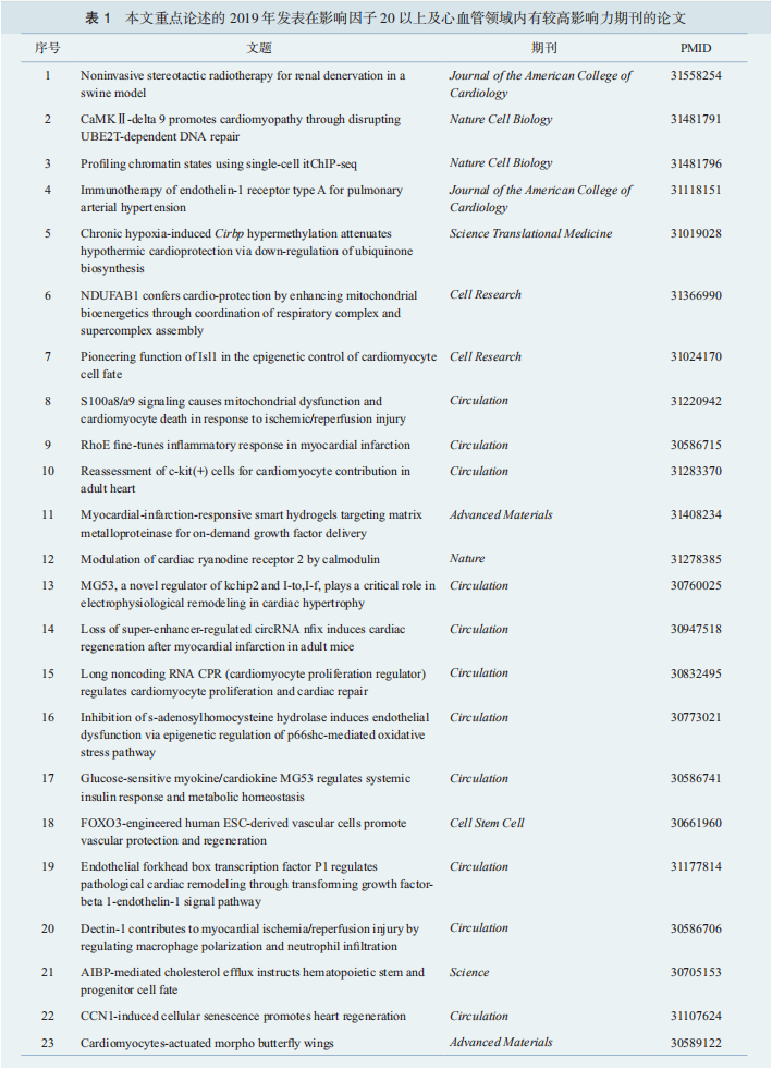
[7] Li Y, Chen B, Yang X, et al. S100a8/a9 signaling causes mitochondrial dysfunction and cardiomyocyte death in response to ischemic/reperfusion injury[J]. Circulation, 2019,140(9):751-764. DOI: 10.1161/CIRCULATIONAHA.118.039262.
[8] Srinivas Bharath MM. Post-translational oxidative modifications of mitochondrial complex I (NADH: ubiquinone oxidoreductase): implications for pathogenesis and therapeutics in human diseases[J]. J Alzheimers Dis, 2017,60(s1):S69-S86. DOI: 10.3233/JAD-170117.
[9] Hou T, Zhang R, Jian C, et al. NDUFAB1 confers cardioprotection by enhancing mitochondrial bioenergetics through coordination of respiratory complex and supercomplex assembly[J]. Cell Res, 2019,29(9):754-766. DOI: 10.1038/s41422-019-0208-x.
[10] Zhang M, Gao H, Liu D, et al. CaMKⅡ-δ9 promotes cardiomyopathy through disrupting UBE2T-dependent DNA repair[J]. Nat Cell Biol, 2019,21(9):1152-1163. DOI: 10.1038/s41556-019-0380-8.
[11] Liu Y, Xing J, Li Y, et al. Chronic hypoxia-induced Cirbp hypermethylation attenuates hypothermic cardioprotection via down-regulation of ubiquinone biosynthesis[J]. Sci Transl Med, 2019,11(489):eaat8406.DOI: 10.1126/scitranslmed.aat8406.
[12] Liu J, Zhuang T, Pi J, et al. Endothelial forkhead box transcription factor P1 regulates pathological cardiac remodeling through transforming growth factor-β1-endothelin-1 signal pathway[J]. Circulation,2019,140(8):665-680. DOI: 10.1161/CIRCULATIONAHA.119.039767.
[13] 张茂,张岩 . 2018 年中国心血管领域主要基础研究回顾 [J/OL]. 中华心血管病杂志 ( 网络版 ),2019,2:e1000019(2019-08-09)[2020-03-18]. http://www.cvjc.org.cn/index.php/Column/columncon/article_id/182. DOI:10.3760/cma.j.issn.2096-1588.2019.1000019.
[14] Frangogiannis NG. Inflammation in cardiac injury, repair and regeneration[J]. Curr Opin Cardiol, 2015, 30(3): 240-245. DOI: 10.1097/HCO.0000000000000158.
[15] Shirazi L F, Bissett J, Romeo F, et al. Role of inflammation in heart failure[J]. Curr Atheroscler Rep, 2017, 19(6): 27. DOI: 10.1007/s11883-017-0660-3.
[16] Marchant DJ, Boyd JH, Lin DC, et al. Inflammation in myocardial diseases[J]. Circ Res, 2012, 110(1): 126-144. DOI:10.1161/CIRCRESAHA.111.243170.
[17] Dai Y, Song J, Li W, et al. RhoE fine-tunes inflammatory response in myocardial infarction[J]. Circulation, 2019,139(9):1185-1198. DOI: 10.1161/CIRCULATIONAHA.118.033700.
[18] Fan Q, Tao R, Zhang H, et al. Dectin-1 contributes to myocardial ischemia/reperfusion injury by regulating macrophage polarization and neutrophil infiltration[J]. Circulation,2019,139(5):663-678. DOI: 10.1161/CIRCULATIONAHA.118.036044.
[19] van Berlo JH, Kanisicak O, Maillet M, et al. C-Kit+ cells minimally contribute cardiomyocytes to the heart[J]. Nature, 2014,509(7500):337-341. DOI: 10.1038/nature13309.
[20] Ellison GM, Vicinanza C, Smith AJ, et al. Adult c-Kit(pos) cardiac stem cells are necessary and sufficient for functional cardiac regeneration and repair[J]. Cell, 2013,154(4):827-842. DOI: 10.1016/j.cell.2013.07.039.
[21] Vicinanza C, Aquila I, Cianflone E, et al. Kitcre knock-in mice fail to fate-map cardiac stem cells[J]. Nature, 2018, 555(7697): E1-E17. DOI:10.1038/nature25771.
[22] He L, Han M, Zhang Z, et al. Reassessment of c-Kit+ cells for cardiomyocyte contribution in adult heart[J]. Circulation, 2019,140(2):164-166. DOI: 10.1161/CIRCULATIONAHA.119.039909.
[23] Guttman M, Amit I, Garber M, et al. Chromatin signature reveals over a thousand highly conserved large non-coding RNAs in mammals[J]. Nature, 2009,458(7235):223-227. DOI: 10.1038/nature07672.
[24] Grote P, Wittler L, Hendrix D, et al. The tissue-specific IncRNA Fendrr is an essential regulator of heart and body wall development in the mouse[J]. Dev Cell, 2013, 24(2): 206-214. DOI:10.1016/j.devcel.2012.12.012.
[25] Wang K, Long B, Zhou LY, et al. CARL lncRNA inhibits anoxia-induced mitochondrial fission and apoptosis in cardiomyocytes by impairing miR-539-dependent PHB2 downregulation[J]. Nat Commun, 2014,5:3596. DOI: 10.1038/ncomms4596.
[26] Kumarswamy R, Bauters C, Volkmann I, et al. Circulating long noncoding RNA, LIPCAR, predicts survival in patients with heart failure[J]. Circ Res, 2014,114(10):1569-1575. DOI: 10.1161/CIRCRESAHA.114.303915.
[27] Ponnusamy M, Liu F, Zhang YH, et al. Long noncoding RNA CPR (cardiomyocyte proliferation regulator) regulates cardiomyocyte proliferation and cardiac repair[J]. Circulation, 2019,139(23):2668-2684. DOI: 10.1161/CIRCULATIONAHA.118.035832.
[28] Yao C, Yu B. Role of long noncoding RNAs and circular RNAs in nerve regeneration[J]. Front Mol Neurosci,2019,12:165. DOI:10.3389/fnmol.2019.00165.
[29] Kristensen LS, Hansen TB, Venø MT, et al. Circular RNAs in cancer: opportunities and challenges in the field[J]. Oncogene,2018,37(5):555-565. DOI:10.1038/onc.2017.361.
[30] Memczak S, Jens M, Elefsinioti A, et al. Circular RNAs are a large class of animal RNAs with regulatory potency[J]. Nature,2013,495(7441):333-338. DOI:10.1038/nature11928.
[31] Werfel S, Nothjunge S, Schwarzmayr T, et al. Characterization of circular RNAs in human, mouse and rat hearts[J]. J Mol Cell Cardiol, 2016,98:103-107. DOI: 10.1016/j.yjmcc.2016.07.007.
[32] Huang S, Li X, Zheng H, et al. Loss of super-enhancer-regulated circRNA nfix induces cardiac regeneration after myocardial infarction in adult mice[J]. Circulation, 2019,139(25):2857-2876. DOI: 10.1161/CIRCULATIONAHA.118.038361.
[33] He S, Sharpless NE. Senescence in health and disease[J]. Cell, 2017,169(6):1000-1011. DOI: 10.1016/j.cell.2017.05.015.
[34] Coppé JP, Desprez PY, Krtolica A, et al. The senescenceassociated secretory phenotype: the dark side of tumor suppression[J]. Annu Rev Pathol, 2010,5:99-118. DOI: 10.1146/annurev-pathol-121808-102144.
[35] Tchkonia T, Zhu Y, van Deursen J, et al. Cellular senescence and the senescent secretory phenotype: therapeutic opportunities[J]. J Clin Invest, 2013,123(3):966-972. DOI: 10.1172/JCI64098.
[36] Feng T, Meng J, Kou S, et al. CCN1-induced cellular senescence promotes heart regeneration[J]. Circulation, 2019,139(21):2495-2498. DOI: 10.1161/CIRCULATIONAHA.119.039530.
[37] Gao R, Liang X, Cheedipudi S, et al. Pioneering function of Isl1 in the epigenetic control of cardiomyocyte cell fate[J]. Cell Res, 2019,29(6):486-501. DOI: 10.1038/s41422-019-0168-1.
[38] Xiao Y, Xia J, Cheng J, et al. Inhibition of S-adenosylhomocysteine hydrolase induces endothelial dysfunction via epigenetic regulation of p66shc-mediated oxidative stress pathway[J]. Circulation, 2019,139(19):2260-2277. DOI: 10.1161/CIRCULATIONAHA.118.036336.
[39] Gu Q, Yang X, Lv J, et al. AIBP-mediated cholesterol efflux instructs hematopoietic stem and progenitor cell fate[J]. Science, 2019,363(6431):1085-1088. DOI: 10.1126/science.aav1749.
[40] Yan P, Li Q, Wang L, et al. FOXO3-engineered human ESCderived vascular cells promote vascular protection and regeneration[J]. Cell Stem Cell, 2019,24(3):447-461. DOI: 10.1016/j.stem.2018.12.002.
[41] Oparil S, Schmieder RE. New approaches in the treatment of hypertension[J]. Circ Res, 2015,116(6):1074-1095. DOI: 10.1161/CIRCRESAHA.116.303603.
[42] Cai X, Yang Y, Shen Y, et al. Noninvasive stereotactic radiotherapy for renal denervation in a swine model[J]. J Am Coll Cardiol, 2019,74(13):1697-1709. DOI: 10.1016/j.jacc.2019.07.053.
[43] Dai Y, Chen X, Song X, et al. Immunotherapy of endothelin-1 receptor type A for pulmonary arterial hypertension[J]. J Am Coll Cardiol, 2019,73(20):2567-2580. DOI: 10.1016/j.jacc.2019.02.067.
[44] Shah PK. Active and passive vaccination for pulmonary arterial hypertension: a novel therapeutic paradigm[J]. J Am Coll Cardiol, 2019,73(20):2581-2583. DOI: 10.1016/j.jacc.2019.02.064.
[45] Ai S, Xiong H, Li CC, et al. Profiling chromatin states using single-cell itChIP-seq[J]. Nat Cell Biol, 2019,21(9):1164-1172. DOI: 10.1038/s41556-019-0383-5.
[46] Fan C, Shi J, Zhuang Y, et al. Myocardial-infarctionresponsive smart hydrogels targeting matrix metalloproteinase for on-demand growth factor delivery[J]. Adv Mater, 2019,31(40):e1902900. DOI: 10.1002/adma.201902900.
[47] Chen Z, Fu F, Yu Y, et al. Cardiomyocytes-actuated morpho butterfly wings[J]. Adv Mater,2019,31(8):e1805431. DOI: 10.1002/adma.201805431.
[48] Zhang Y, Wu HK, Lv F, et al. MG53: biological function and potential as a therapeutic target[J]. Mol Pharmacol, 2017,92(3):211-218. DOI: 10.1124/mol.117.108241.
[49] Zhang Y, Wu HK, Lv FX, et al. MG53 is a double-edged sword for human diseases[J]. Sheng Li Xue Bao, 2016,68(4):505-516.
[50] Cao CM, Zhang Y, Weisleder N, et al. MG53 constitutes a primary determinant of cardiac ischemic preconditioning[J]. Circulation, 2010,121(23):2565-2574. DOI: 10.1161/CIRCULATIONAHA.110.954628.
[51] Cai C, Masumiya H, Weisleder N, et al. MG53 nucleates assembly of cell membrane repair machinery[J]. Nat Cell Biol, 2009,11(1):56-64. DOI: 10.1038/ncb1812.
[52] Jia Y, Chen K, Lin P, et al. Treatment of acute lung injury by targeting MG53-mediated cell membrane repair[J]. Nat Commun, 2014,5:4387. DOI: 10.1038/ncomms5387.
[53] Kim SC, Kellett T, Wang S, et al. Modulation of Trim72 alters the repair capacity of lung epithelial cells[J]. Am J Physiol Lung Cell Mol Physiol,2014,307:L449-L459. DOI:10.1152/ajplung.00172.2014.
[54] Duann P, Li H, Lin P, et al. MG53-mediated cell membrane repair protects against acute kidney injury[J]. Sci Transl Med, 2015,7(279):279ra36. DOI: 10.1126/scitranslmed.3010755.
[55] Weisleder N, Takizawa N, Lin P, et al. Recombinant MG53 protein modulates therapeutic cell membrane repair in treatment of muscular dystrophy[J]. Sci Transl Med, 2012,4(139):139ra85. DOI: 10.1126/scitranslmed.3003921.
[56] Yao Y, Zhang B, Zhu H, et al. MG53 permeates through bloodbrain barrier to protect ischemic brain injury[J]. Oncotarget, 2016,7(16):22474-22485. DOI: 10.18632/oncotarget.7965.
[57] Zhang Y, Lv F, Jin L, et al. MG53 participates in ischaemic postconditioning through the RISK signalling pathway[J]. Cardiovasc Res, 2011,91(1):108-115. DOI: 10.1093/cvr/cvr029.
[58] Yi JS, Park JS, Ham YM, et al. MG53-induced IRS-1 ubiquitination negatively regulates skeletal myogenesis and insulin signalling[J]. Nat Commun, 2013,4:2354. DOI: 10.1038/ncomms3354.
[59] Song R, Peng W, Zhang Y, et al. Central role of E3 ubiquitin ligase MG53 in insulin resistance and metabolic disorders[J]. Nature, 2013,494(7437):375-379. DOI: 10.1038/nature11834.
[60] Liu F, Song R, Feng Y, et al. Upregulation of MG53 induces diabetic cardiomyopathy through transcriptional activation of peroxisome proliferation-activated receptor α[J]. Circulation, 2015,131(9):795-804. DOI: 10.1161/CIRCULATIONAHA.114.012285.
[61] Grundy SM. Metabolic syndrome: connecting and reconciling cardiovascular and diabetes worlds[J]. J Am Coll Cardiol, 2006,47(6):1093-1100. DOI: 10.1016/j.jacc.2005.11.046.
[62] Wu HK, Zhang Y, Cao CM, et al. Glucose-sensitive myokine/cardiokine MG53 regulates systemic insulin response and metabolic homeostasis[J].Circulation,2019,139(7):901-914. DOI:10.1161/CIRCULATIONAHA.118.037216.
[63] Liu W, Wang G,Zhang C,et al. MG53, anovel regulator of KChIP2 and Ito,f, plays a critical role in electrophysiological remodeling in cardiac hypertrophy[J]. Circulation, 2019,139(18):2142-2156. DOI: 10.1161/CIRCULATIONAHA.118.029413.
[64] Patel SP, Campbell DL. Transient outward potassium current, ‘Ito’, phenotypes in the mammalian left ventricle: underlying molecular, cellular and biophysical mechanisms[J]. J Physiol, 2005,569(Pt 1):7-39. DOI: 10.1113/jphysiol.2005.086223.
[65] Dong M, Yan S, Chen Y, et al. Role of the transient outward current in regulating mechanical properties of canine ventricular myocytes[J]. J Cardiovasc Electrophysiol, 2010,21(6):697-703. DOI: 10.1111/j.1540-8167.2009.01708.x.
[66] Thomsen MB, Sosunov EA, Anyukhovsky EP, et al. Deleting the accessory subunit KChIP2 results in loss of I(to,f) and increased I(K,slow) that maintains normal action potential configuration[J]. Heart Rhythm, 2009,6(3):370-377. DOI: 10.1016/j.hrthm.2008.11.023.
[67] Gong D, Chi X, Wei J, et al. Modulation of cardiac ryanodine receptor 2 by calmodulin[J]. Nature, 2019,572(7769):347-351. DOI: 10.1038/s41586-019-1377-y.